It is a common notion that sophisticated renderings of anatomy began in the Renaissance, such as Leonardo da Vinci’s drawings of a dissected womb, when in fact, complex drawings of organs existed over a century before. Rare examples of such are found in MS Ashmole 399, a medical compendium created in England in the late-thirteenth century (see below for a quick ‘tour’ of the manuscript). The manuscript, consisting of seventy-three leaves of vellum and measuring 270 x 195 mm, contains a set of anatomical drawings produced in the last quarter of the thirteenth century, inserted into the compendium circa 1298, according to palaeographical analysis. In the decades following, Latin medical tracts were written into the spaces surrounding the anatomical drawings.
A selection of these drawings make up what is now referred to as the 'Nine-figure series': the earliest set of anatomical drawings in Western Europe to have survived. These anatomical images together give the viewer a tour of the human anatomy, including:
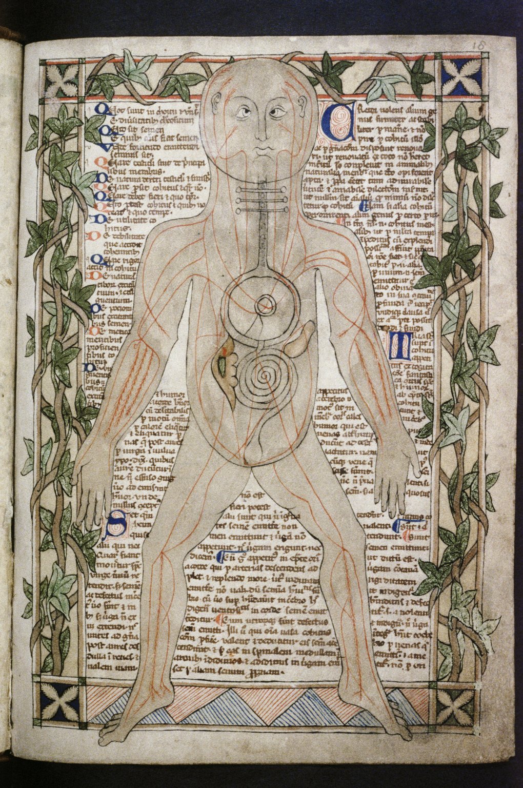
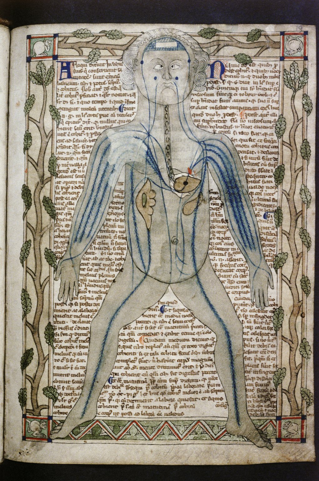
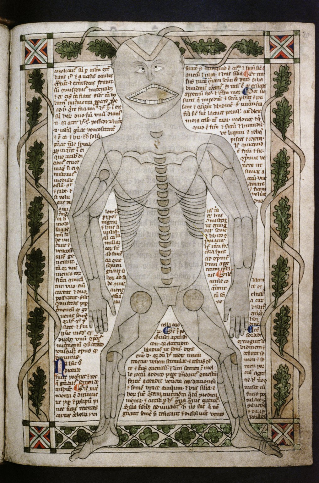
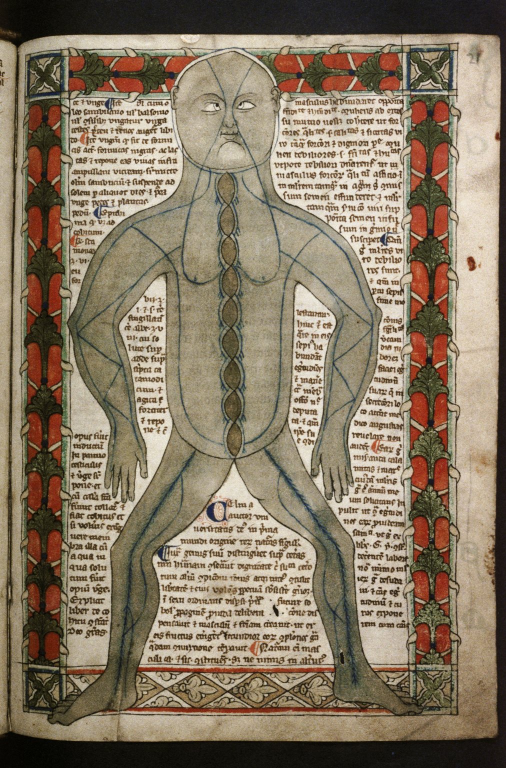
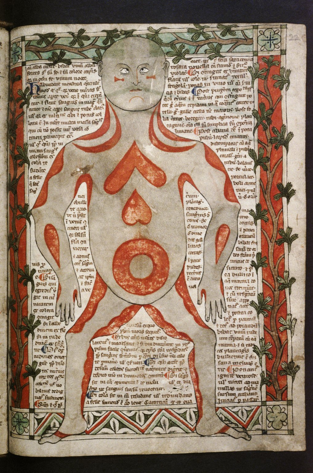
·A set of five images which show the different systems of the body (the veins, skeleton, arteries, skeleton, nerves, and muscles) within a template of a human figure (folios 18r-22r, above). With the exception of the first (vein) man, each system within the human figure is described on its opposing leaf in black textualis with an initial illuminated with gold-leaf decoration. These have historically been referred to as the Fünfbilderserie, or 'five-figure series', as they are found together in many instances without the following drawings;
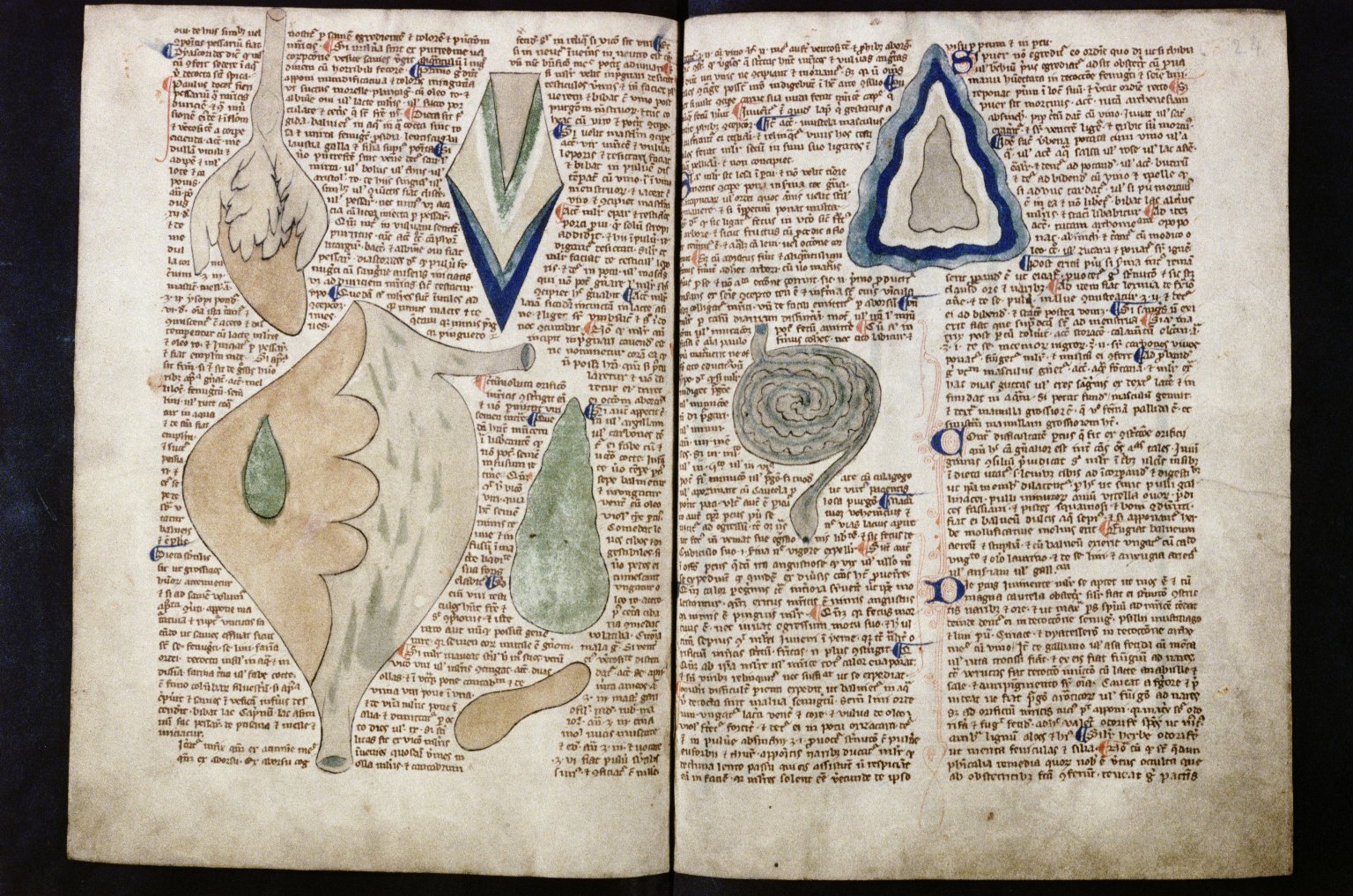
·A group of naturalistic organ renderings including the kidneys, liver, stomach and heart (folios 23v-24r, above);
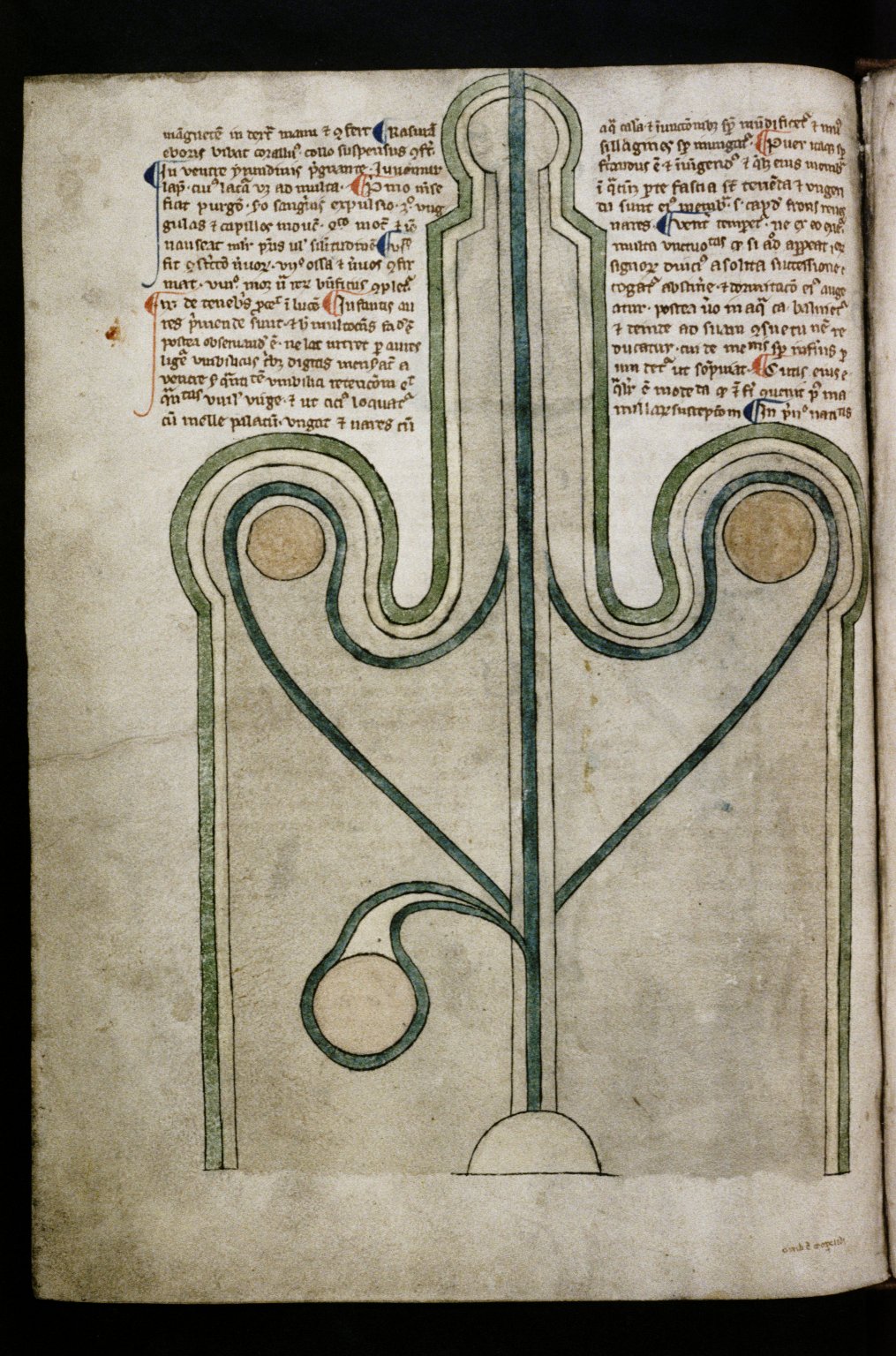
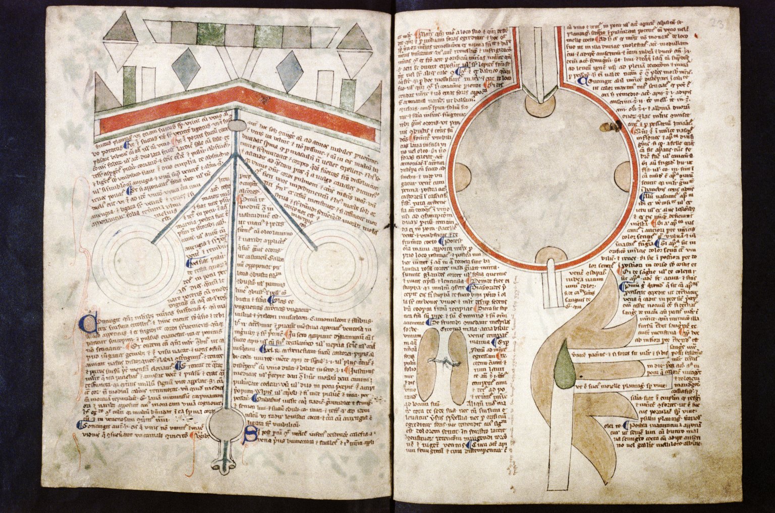
·Schematic diagrams of the penis (fol. 24v, above), the eyes and brain (fol. 22v, above), the stomach (fol. 23r, above), and, most curiously, a diagram of the uterus (fol. 13v, below). Ten foetus-in-utero images (folios 14v- 15r, below) also immediately succeed the uterus diagram, but these are not always associated with the nine-figure series.
Despite the distinct ways in which the organs are represented, their visual analysis suggests that they were drawn by one artist. For example, they all share the same thick black contour and thin washes of colour. Only a handful of examples of the nine-figure series survive. The most similar copy is found in a late-twelfth century libellus which forms part of Cambridge, Gonville and Caius College MS 190/223 and from which it has been suggested that the Bodleian version was copied. This image set is rare now, but there is no way of knowing just how many would have existed in the Middle Ages, especially if they were incorporated into manuscripts with a practical, medical use. The organ drawings of Ashmole 399 have an overwhelming amount to tell us about the study of anatomy in the Middle Ages, as well as the role of images in the learning and practice of medicine. This article therefore focuses on just one of these images, which has captivated historians of medicine and art historians alike for over a century: the diagram of the uterus.
The Uterus Diagram
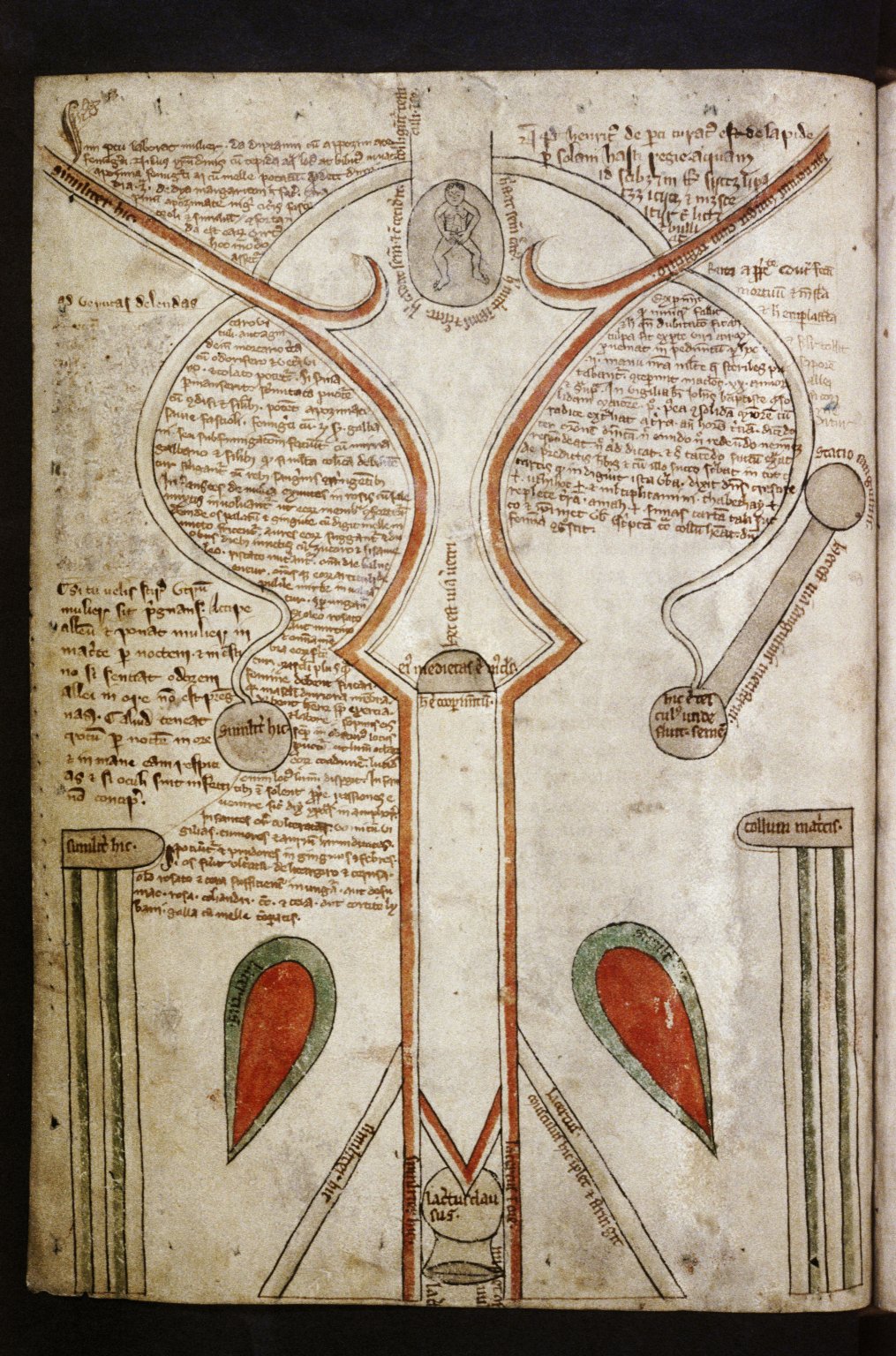
The depiction of the uterus is curiously schematic, its architectural appearance emphasised by the fluted column-like structures with which it is framed. Upon closer inspection, it becomes clear that the image is concerned with conception and generation: the physiological purpose of the organ. At the top of the vaginal canal at the centre of the image is the plug (cooperminentum) of the womb, above which it is written 'this is the way of the male private parts' (hec est via veretri), to show where the penis is inserted for conception to take place. Above this is the resulting embryo contained within an oval.
There are inherent issues, practical and social, in the depiction of the female genitalia. Practically, the female reproductive system is particularly difficult to represent as not only is it hidden within the body, but it is also ever-changing in a monthly cycle. Socially, there are implications derivative of contemporary perceptions of sexual difference, in which the female body was often perceived to have an aura of shame. This makes the image type of the ‘schematic uterus’ particularly revealing of contemporary issues of dissection and gender: central concerns to historians of medicine.
The diagram provokes many questions as to how it was made and used: How was the appearance of the female reproductive system known at a time when there were social taboos surrounding human dissection? Why was the organ rendered in a schematic form? Was it intended as a realistic portrayal of the appearance of the uterus, or was it trying to show something else? How was the image intended to be used, and did it have a practical medical function? What can the image tell us about medieval attitudes towards the female body? Through the image's analysis and contextualisation, we can explore the various potential influences which informed its rendering, including: dissection, Islamic medicine, medical texts and the perception of women during the Middle Ages.
Dissection
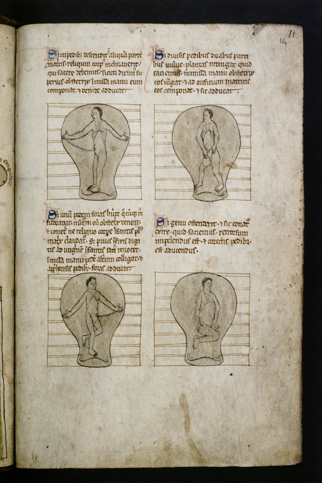
Initially striking about the image is how modern it looks. Depictions of the female anatomy were not entirely rare in the Middle Ages, but in the majority of the surviving images the womb is shown as pregnant and often light-bulb shaped. Examples of this are found immediately following the Ashmole uterus diagram, where ten outlines of wombs contain adult-looking foetuses in various disco-reminiscent poses. Yet this differs entirely from what a viewer today would expect of a uterus diagram, such as those found in biology text books or tampon instructions which show a cross section of the organ and therefore its internal features. The purpose of the schematic uterus is not to express its shape during pregnancy, as in the more common medieval depictions of the organ, but to illustrate the different components of the female reproductive system and how they work.
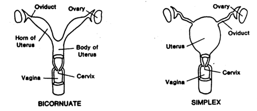
In order to represent its appearance, the artist of the original diagram must have worked from either first-hand observation of the organ, or from a textual description of it. The image is therefore revealing of medieval practices of medical learning and dissection. Particular features of the depicted organ, for example, indicate that the artist (or the writer who described it) did not draw from the uterus of a human, but from an animal. Contemporary accounts of pig anatomy describe their uterine horns, a feature of bicornate uteri which are found in many mammals, such as cows or pigs. This feature is visible in fol. 13v yet is not present in the human, i.e. simplex, uterus (see above; for a three-dimensional rendering of the simplex uterus, see here). This accurately reflects contemporary medical practice; although there is no conclusive evidence that human dissection was strictly illegal in thirteenth-century England, it was rare that a human would have been subject to it. The question of whether humans were routinely dissected in the Middle Ages is, however, a contentious one. The dissection of pregnant women in particular would remain an issue for centuries to come, as shown by the controversy surrounding the eighteenth-century engravings of the womb in William Hunt's Anatomia Uteri Humani Gravidi Tabulis Illustrata. The insides of animal's bodies, on the other hand, whether observed for anatomical study or during culinary preparation, were not a mystery.
Islamic Medicine
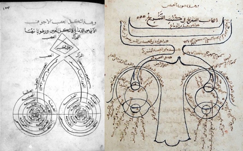
Where and when exactly the original uterus diagram was made is still uncertain and it is possible that it, as well as the entire nine-figure series, has an Islamic origin. This is supported by the schematic representation of the eyes and brain on fol. 22v. The eyes are represented by concentric circles and feed images of the external world through the blue line into the brain, which consists of geometrical shapes representing neurological function. Similar Islamic representations of the eyes and brain can be found in the Jawāmi', Galen’s lost treatise on eye diseases (above left, dated 1431) and its predecessor is found in a copy of Ibn Al-Haytham’s treatise on optics (above right, dated 1083). Like these Islamic images, the schematic drawings of Ashmole 399 render organic structures into a geometric form, representing both their physiognomic appearance as well as their physiological function.
Text
The ways in which text is applied to the uterus diagram, both the short captions and the longer texts, indicate how the image was received. Treatises are written around the other schematic diagrams of the penis, eyes and brain, and stomach in a careful and neat script, whereas the schematic uterus is written upon extensively and in multiple hands. The uterus diagram is also the only image whose individual components are labelled.
The longer texts surrounding the uterus are later additions. They consist of at least two different hands showing that they have been added over time, creating a collection of scribbled practical remedies which use the diagram as their base. This includes prescriptions for the use of pregnant and parturient women. The practical content of the text is reflected in its untidiness; it is barely legible, full of abbreviations, inconsistent, and some of the text on the left of the diagram has been scratched out in an untidy manner. The same hand appears to have outlined segments of the diagram in a lighter brown ink and drawn the horizontally-gridded boxes within the light-bulb wombs on the following pages.
But what can this tell us about how the diagram was used and perceived in the Middle Ages? Monica Green, in her study of medical texts concerning women in the Middle Ages, showed that five of the eight of Ashmole 399’s texts are copied from another medical miscellany (MS Oxford, Bodleian Library, Pembroke 21) in which gynaecology and embryology are consciously separated. She thus argued that the author of the Ashmole 399 treatises created a trilogy of texts relating to human generation: Constantine the African’s De Coitu, the pseudo-Galenic De Spermate and the Trotula, a twelfth-century compendium of women's medicine. At a later point, another hand added Constantine the African’s chapters referring to the generative organs, the De Genecia, directly after the foetus-in utero images. By altering the grouping of texts, the later scribes of Ashmole 399 consciously juxtaposed gynaecological and generational texts, transforming the manuscript into what Green referred to as ‘a veritable summa on generation’, reflective of the changing perception of women in thirteenth-century medicine. This is emphasised by the texts inserted around the uterus diagram, which contain recipes for aiding conception and childbirth.
The female body in the Middle Ages
Many historians have suggested reasons for the emergence of a women’s medicine at this time. Katherine Park has written extensively on the medieval perception of the female body, arguing that its anatomical significance derives from its classification as the ‘non-generic body’ opposite the generic male body. The transformation of attitudes concerning the female body, from curing their diseases to understanding their secrets, was shown by the change in vocabulary in medical tracts. In the thirteenth century, the female genitals became more commonly referred to as loca secreta (hidden places) and pudibunda (shameful parts). In the schematic uterus of Ashmole 399, we can see this desire to understand and convey the inner-workings of the female reproductive system.
Like the majority of those who made anatomical drawings, the maker of the diagram was faced with the challenge of capturing the appearance and function of a complicated three-dimensional organ upon a two-dimensional plane. Instead of using light and shade to capture the realistic appearance of the organ, like the aforementioned eighteenth-century engravings of the womb, the Ashmole 399 artist rendered it in a geometrical form which illustrated how its different components work together to perform generation. The uterus here is not lifeless flesh, but a working mechanism, the diagram its blueprint. Just as the lift-the-flap anatomical books which came a couple of centuries later, the schematic uterus allowed the viewer to peel back the outer layer of the light-bulb womb, to reveal the secrets of women and the miracle of creation.
Further Reading
Green, M. H., From “Diseases of Women” to “Secrets of Women”: The Transformation of Gynaecological Literature in the Later Middle Ages’ Journal of Medieval and Early Modern Studies, 30:1, (Winter 2000), 5-39.
Jones, P., ‘Image, Word and Medicine in the Middle Ages,’ in Vizualising Medieval Medicine and Natural History 1200-1500, ed. by J. A. Givens, K. M. Reeds and A. Touwade (Ashgate, 2006), pp. 1-24.
McCall, T., ‘Reliquam Dicit Pictura: Text and Image in a Twelfth-Century Illustrated Anatomical Manual (Gonville and Caius College, Cambridge, MS 190/223),’ Transactions of the Cambridge Bibliographical Society, Vol. 16, No. 1 (2016), 1-22.
- ‘Illuminating the Interior: The Illustrations of the Nine Systems of the Body and Anatomical Knowledge in Medieval Europe’ (PhD dissertation, University of Cambridge, 2017)
Park, K., Secrets of Women: Gender, Generation and the Origins of Human Dissection (New York, 2006)
Savage-Smith, E., ‘Anatomical Illustration in Arabic Manuscripts,’ from Arab Painting: Text and Image in Illustrated Arabic Manuscripts, ed. by Anna Contadini (Leiden, 2007), 147-59.
Whittington, K., ‘The Cruciform Womb: Process, Symbol and Salvation in Bodleian Library MS. Ashmole 399,’ Different Visions: A Journal of New Perspectives on Medieval Art, Issue 1 (2008), 1-24.